Could we be targeting Alzheimer’s wrong? NEOMED researcher explores a new approach
June 4, 2021
The sixth leading cause of death in the United States, Alzheimer’s disease (AD) is the only one in the top ten that cannot be prevented, slowed, or cured.
Many scientists in the area of Alzheimer’s research focus on finding a way to prevent cell death — believed to be the end stage event in a neuron’s decline. But this may be one of the reasons all of the clinical trials of AD therapies have failed, says Samuel Crish, Ph.D., associate professor in the Department of Pharmaceutical Sciences. After all, if you keep a neuron from dying but it can’t communicate, you will still have the same symptoms.
Not long ago, Dr. Crish took a mental step back and asked himself this: What if we have been targeting the wrong process?
Supported by new funding from the Templeton Medical Research Foundation, Dr. Crish’s lab will investigate the concept of neuronal dysfunction as a driver of degeneration and target for intervention.
“Our research investigates the possibility of restoring function to “sick” neurons in Alzheimer’s disease. This may result in much-needed therapies that stop the progression of neurodegeneration and may actually reverse some clinical symptoms,” Dr. Crish explains.
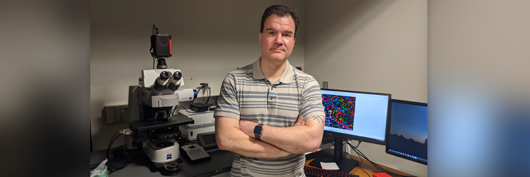
Developing disease-modifying therapies for brain health
Alzheimer’s disease is a chronic neurodegenerative condition that gradually destroys a person’s cognitive abilities – learning and memory, reasoning, and planning – and leads to death within five to 12 years after diagnosis. Functioning in the Neurodegenerative Disease and Aging Research Focus Area, Dr. Crish’s laboratory focuses on developing disease-modifying therapies to prevent, slow, stop, or even reverse damage to the brain.
The Alzheimer’s disease research Dr. Crish leads is an outgrowth of the research he was doing in glaucoma – another age-related chronic neurodegeneration – when he first came to NEOMED in 2010.
Links and early clues: Alzheimer’s and glaucoma
“We discovered that neurotoxic proteins associated with Alzheimer’s disease, namely amyloid-beta and tau, play a role in retinal degeneration in glaucoma. That got us thinking that there may be other similarities between the two conditions, so we started investigating Alzheimer’s disease in 2016 and it has since become a major topic of our research,” says Dr. Crish.
“Our lab looks at these diseases differently that most investigators. While most AD and glaucoma research concentrate on neuronal death as the cause of clinical deficits, we have found that neurons lose the ability to communicate long before they die. In this case, that means the eye stops sending visual information to the brain. While the standard thinking is that this happens because retinal cells are lost, we found that many of these cells are still present; they just stop signaling other neurons. This creates an exciting therapeutic window where we may be able to restore function to these ‘sick’ cells – stopping the progression of degeneration and possibly restoring some lost capabilities.”
Hope for treatment
The visual deficits that are so common in AD have a substantial impact on quality of life, as any patient could attest.
Dr. Crish’s lab has found that nonfunctional retinal cells in glaucoma can be “rescued” by treatment with a multiple sclerosis drug, fingolimod (previous work also supported by the Templeton Foundation). “Given the similarities between AD and glaucoma, we believe that treatment with fingolimod or similar agents may restore damaged neurons’ function in AD,” he says.
The importance of his lab’s work is threefold, says Dr. Crish.
First, it identifies restoring neuronal function as a target for intervention.
Second, it supports the idea of retinal changes as a much-needed biomarker. That concept is really significant, because changes in the eye can precede brain pathology by a decade or more, allowing for identification of a person’s risk of developing AD or the stage to which the disease has progressed. And, Dr. Crish adds, the retina can be non-invasively imaged by a person’s normal eye doctor, not an expensive specialist.
Finally, his lab’s findings in the visual system can be extrapolated to the brain. In other words, therapies that work in the visual system will also be likely to work in the parts of the brain responsible for cognitive function. With this latest grant funding, Dr. Crish and his team can work toward these goals.